Oxygen-Sensitive Remodeling of Central Carbon Metabolism by Archaic elF5B

Author: J.J. David Ho et al.
Date: January 2018

Oxygen-Sensitive Remodeling of Central Carbon Metabolism by Archaic eIF5B

eIF5B is a GTPase eukaryotic translation initiation factor found on ribosomes which positions the initiation methionine tRNA on start codons of respective mRNA to ensure translation accurately initiates. It is speculated that aerobic eukarya retained eIF5B to remodel anaerobic pathways during episodes of oxygen deficiency.

The authors developed a method with the capability to generate an architectural blueprint of biologically active cellular translational machineries, termed MATRIX. This method combines metabolic pulse labeling, ribosome density fractionation and high-throughput mass spectrometry. MATRIX was employed to compare the protein abundance in polysome fractions (active translation) to that of free fractions (translationally disengaged) in both normoxic and hypoxic cells, ultimately proving that eIF5B concentrates in hypoxic translating ribosomes.

Read more: Oxygen-Sensitive Remodeling of Central Carbon Metabolism by Archaic elF5B

Poldip2 is an oxygen-sensitive protein that controls PDH and αKGDH lipoylation and activation to support metabolic adaptation in hypoxia and cancer

Author: Felipe Paredes et al.
Date: January 2018
Poldip2 is an oxygen-sensitive protein that controls PDH and αKGDH lipoylation and activation to support metabolic adaptation in hypoxia and cancer

In this paper, the authors show that poldip2 governs the critical mechanism linking clp, ACSM1, and protein lipoylation; thus, regulating mitochondria function.

Lipoylation of two key enzymes in the TCA cycle, PDH and αKGDH, is a dynamically regulated process that is inhibited under hypoxia and in cancer cells, resulting in restricted mitochondria function. Poldip2, a ubiquitously expressed protein, regulates the lipoylation of both pyruvate and αKDH subunits via regulation of the clp-protease complex (CLPX ) and deregulation of the lipoate-activating enzyme required for lipoylation in mammalian cells, Ac-CoA synthetase (ACSM1). In Poldip2-defficient cells, repressed mitochondria function induces the stabilization of HIF-1α, thereby reprogramming cellular metabolism to resemble hypoxic/cancer cell adaption. Additionally, Poldip2 is down-regulated in hypoxic environments, further stabilizing HIF-1α while also inducing the expression of PDH kinase (PDK), consequently inhibiting lipoylation.

Read more: Poldip2 is an oxygen-sensitive protein that controls PDH and αKGDH lipoylation and activation to...

Molecular Targeting of Hypoxia in Radiotherapy

Most solid tumors exhibit areas of both chronic and acute hypoxia, all of them evolving dynamically as a function of cellular growth, vascularization, oxygen consuming metabolism and therapy response. Tumor hypoxia, generally far below 1% oxygen, correlates with increased recurrence rates and decreased survival rates in most cancers, so the recent review by HypOxystation users Rey et al. describing “Molecular Targeting of Hypoxia in Radiotherapy”  gives a valuable overview of the mechanisms cancer cells have developed to respond to hypoxia.

Dr. Rey of the Princess Margaret Cancer Centre in Toronto, Canada, and his co-authors Luana Schito, Marianne Koritzinsky and Brad Wouters have contributed vastly to our knowledge about the cellular response to hypoxia in the context of tumor behavior. Since 2009, they have acquired 4 HypOxystations for their lab, in order to culture cells under conditions which authentically mimic the physiological environment of cancer. The HypOxystation provides a closed workstation format for rigorous control of oxygen, CO2, temperature and humidity, facilitating accurate regulation of cell culture conditions as the in vivo tumor situation is simulated. An extensive list of publications from our HypOxystation users spans the whole breadth of research into metabolic, epigenetic and therapeutic implications of hypoxia.

Read more: Molecular Targeting of Hypoxia in Radiotherapy

Tumor Metabolism and Hypoxia

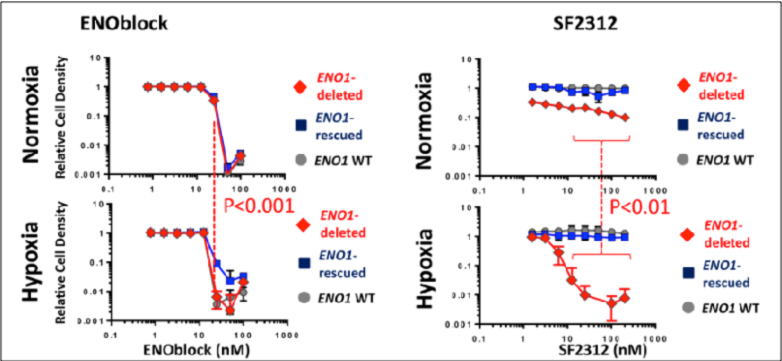
1. HypOxystation users Satani et al. examined the efficacy of ENOblock, a promising new drug for the treatment of cancer and diabetes which was thought to inhibit the glycolytic enzyme enolase. In their recent paper “ENOblock Does Not Inhibit the Activity of the Glycolytic Enzyme Enolase”, the group at MD Anderson Cancer Center in Houston used data from X-ray structures, Cellular thermal shift assays and mutational analyses to show that while the biological effects of the cell permeable ENOblock were reproduced in glioma cells cultured under hypoxia (0.1%), the efficacy of the drug must involve other mechanisms than the previously reported direct inhibition of enolase activity.
tumor1
From: Satani et al. (2016) “ENOblock Does Not Inhibit the Activity of the Glycolytic Enzyme Enolase” PLoS ONE 11(12):e0168739. doi:10.1371

Read more: Tumor Metabolism and Hypoxia

Adaptation to Stressors by Systemic Amyloidogenesis

Cells facing environmental threats have developed numerous coping mechanisms, and HypOxystation users Tim Audas and Stephen Lee have uncovered a fascinating new cellular strategy to remain viable under stress and restore homeostasis when the crisis ends. In their recent paper “Adaptation to Stressors by Systemic Protein Amyloidogenesis”, they describe a physiological process of amyloidogenesis which cells activate under stress conditions, such as hypoxia and acidosis, to remove copious amounts of heterogeneous proteins from circulation, enabling cells to survive in a dormant state. This discovery expands our current view of amyloids as a rare and pathological phenomenon associated with neuropathies such as Alzheimer’s and Parkinson’s diseases, and exposes a novel post-translational, regulatory form of protein organization.

Using a combination of Congo red staining, proteinase K digestion, and OC antibody detection on cells exposed to a variety of stimuli, Audas et al. were able to identify nuclear foci consisting of immobilized, insoluble protein in a crossed β-sheet conformation which they named A-Bodies. In amyloidogenic proteins such as VHL and RNF8, an ACM (amyloid-converting motif) containing arginine and histidine was identified as essential for capture specifically in the A-bodies; a similar motif was also identified in the pathological β-amyloid associated with Alzheimer’s disease. Upon environmental insult, the ACM interacts with ribosomal intergenic spacer RNA (rIGSRNA) to concentrate the proteins and trigger their polymerization in the A-bodies allowing the cells to enter a dormant state.

Read more: Adaptation to Stressors by Systemic Amyloidogenesis