

Author: Giulia Cheloni, et al
Date: June 2017
The Leukemic Stem Cell Niche: Adaptation to "Hypoxia" Versus Oncogene Addiction
Hematopoietic stem cells (HSC) are responsible for constantly maintaining and replenishing the supply of new blood and immune cells. They give rise to both lymphoid and myeloid progenitor cells, which then proceed to differentiate down their respective paths to form various specialized cells such as erythrocytes, macrophages, B and T cells, to name a few. Within the body, HSCs are found to reside in special locations termed “stem cell niches.” Stem cell niches (SCN) are sites in bone marrow dedicated to the long term maintenance of hematopoietic stem cells. HSC’s proliferate within SCN environments without losing stem cell potential i.e. they partake in self-renewal without differentiating – a defining characteristic of stem cells.
Unfortunately, like all other regulated cell cycles, the renewal and differentiation of HSC’s can succumb to replication errors which gives rise to many forms of cancer, namely those within the leukemia and lymphoma families. Leukemia diseases specifically, arise when myeloid progenitor cells become malignant aberrant cells naturally called leukemic stem cells (LSC). In this paper, Cheloni et al. primarily focused on chronic myeloid leukemia (CML). In CML, a translocation of DNA occurs between chromosomes 9 and 22, resulting in the production of the gene BCR-Abl which causes CML cells to expand and replicate.
One of the defining characteristics of stem cell niches (SCN) is their incredibly low oxygen concentration. Physiological tissue concentrations of oxygen generally range from 0.1 – 5%, depending on the tissue. SCNs are found at the far left side of this scale, close to about 0.1% O2. While this environment is considered “normoxic” for HSCs, most differentiated adult cells, including leukemic stem cells, do not thrive within these niches. As a result of the incredibly low oxygen concentrations, ATP-dependent protein production is greatly reduced and all resources are allocated to only producing molecules such as VEGF, PDGF, and HIF-α which are essential for survival in hypoxic environments. By metabolically adapting to produce only essential molecules, other signaling molecules, i.e. oncogenic cytokines in the case of LSCs, effectively become silenced.
With the above information in mind, it’s important to note that leukemia cell lines are highly heterogeneous populations comprising of a full spectrum of functional phenotypes. Unique LSCs with metabolic variations can function independently from oncogenic signaling and exploit the SCN as a valid residence. This independency from oncogenes is critical to their survival since most oncogene signaling-addicted LSCs will suffer from the prevalence of pro-apoptosis over pro-survival stimuli upon withdrawal of the oncogenic signals (termed oncogenic shock).
The authors hypothesized that suppression of the BCR-Abl oncogene is likely a key positive regulator of LSC adaptation and survival within “hypoxic” SCNs. The phrase “adaptation to hypoxia” is commonly used without defining the conditions enabling such actions to occur. To withstand the SCN environment, cells need three things: HIF-1α up-regulation, enhanced glycolysis and reduced mitochondrial/respiratory activity. To analyze the various mechanisms and responses that CML cells demonstrate within SCNs, the authors studied correlations between varied oxygen and glucose concentrations with the amount of BCR-Abl produced. All tests were performed using two human CML cell lines, K562 and KCL22. Testing conditions were precisely controlled to mimic the SCN environment as close as possible. Using a HypOxystation supplied by Don Whitely Scientific, a water-saturated atmosphere comprising of 0.1% O2, 94.9% N2 and 5% CO2 was generated and maintained.
The authors proved that CML cells in 0.1% O2 had greatly decreased BCR-Abl output and also found that the decrease in BCR-Abl was correlated with a decrease in downstream phosphorylated CRLK (a major substrate for BCR-Abl proteins). Additionally, they confirmed that glucose consumption & BCR-Abl protein suppression was positively correlated since the concentration of glucose decreased as the concentration of protein present in the culture media also decreased. Almost undetectable concentrations of BCR-Abl were observed in cultures of K562 and KCL22 within 3 and 4 days, respectively.
An important finding was that CML cells residing within a SCN maintained stem cell potential while the oncogenic proteins responsible for the disease were suppressed. Suppression for BCR-Abl was found to occur only when glucose shortage compliments that of oxygen. A crucial by-product of SCN-adaptation is that LSCs develop a refractoriness to tyrosine kinase inhibitors (TKi), the therapeutic drugs used to target the enzymatic activity of BCR-Abl. This characteristic is what allows the long term maintenance of therapy-resistant LSCs-resulting in minimal residual disease (MRD). Minimal residual disease (MRD) is associated with leukemic cancers to describe a condition where trace amounts of cells remain and are the primary cause of relapse for patients in remission.
The research conducted and documented by Cheloni et al. has provided great insight into several key regulatory mechanisms associated with leukemic stem cells as well as an explanation for their notorious reputation for having high relapse rates. Additionally, they established that the triggering of oncogene suppression associated with CML is due to severe energy restriction rather than simply the “adaptation to hypoxia.”
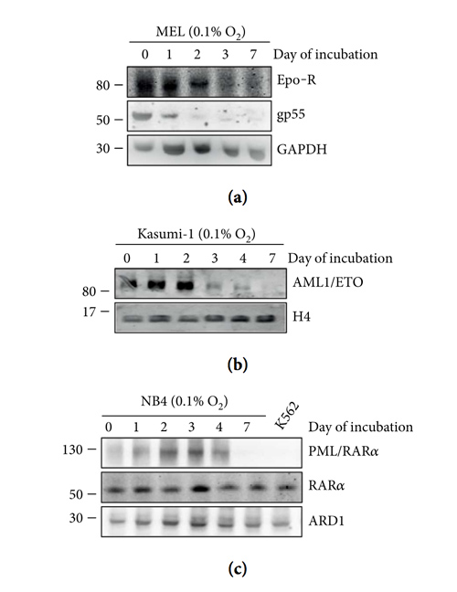
Figure 2: Suppression of oncogenic proteins driving non-CML blood neoplasias in the course of cell “adaptation to hypoxia.” MEL (a), Kasumi-1 (b), or NB4 (c) cells were incubated in atmosphere at 0.1% O2 and lysed at the indicated times, and total cell lysates were subjected to immunoblotting with the indicated antibodies. GAPDH, H4, or ARD1 were detected to verify loading equalization. Migration of molecular weight markers is indicated on the left (kDa). For each cell population, one out of three independent experiments with similar outcome is shown.
